
AI system surpasses radiologists in spotting pancreatic cancer on standard CT scans
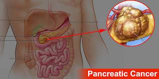

A new observational study has found that an artificial intelligence (AI) model can identify pancreatic ductal adenocarcinoma (PDAC) on routine CT imaging more accurately than human radiologists.
The research team, led by Natalia Alves, MSc, from Radboud University Medical Center in Nijmegen, Netherlands, evaluated the tool in a test group of 1,130 patients. The AI achieved an area under the receiver operating characteristic curve (AUROC) of 0.92 (95% CI 0.90–0.93), along with 85.7% sensitivity and 83.5% specificity at the optimal threshold.
In a secondary analysis of 391 cases, the algorithm delivered not only non-inferior (P<0.0001) but also superior (P=0.001) performance compared with a group of participating radiologists. The clinicians recorded an AUROC of 0.88 (95% CI 0.85–0.91), while the AI once again reached 0.92 (95% CI 0.89–0.94), according to results published in Lancet Oncology. At equivalent sensitivity, the AI system reduced false-positive results by 38% — 85 compared with the radiologists’ 138.
According to the authors, the PANORAMA study is the first large-scale, paired, international diagnostic accuracy investigation that directly compares radiologist performance with a standalone AI model using contrast-enhanced CT scans and a FAIR (findable, accessible, interoperable, and reusable) reference dataset. They concluded that AI trained on broad, diverse patient populations can outperform the average radiologist in PDAC detection, paving the way for future regulatory discussions and real-world trials.
In a commentary accompanying the publication, Misha Luyer, MD, PhD, of Catharina Hospital Eindhoven, described the findings as a major step forward at a time when many AI systems have yet to demonstrate consistent clinical value. He noted that improved early detection is crucial, given the challenges of diagnosing pancreatic cancer at an early stage and the variation in radiology expertise among institutions. Luyer also highlighted that the system’s reduction of false alarms may help limit unnecessary testing and patient stress.
The PANORAMA research consisted of two components: an international AI “grand challenge” and a multi-reader, multi-case study. During the grand challenge, developers worldwide produced algorithms using a publicly available multicenter dataset. Their models were then tested in a controlled, masked setting using predefined metrics.
The observer study involved 68 radiologists from 40 hospitals across 12 countries, all experienced in abdominal CT interpretation.
Overall, the study analyzed 3,440 patient cases (median age 67; 56% male) from five major centers and two public databases. The training set included 2,224 cases, while 86 were used for model tuning. The blinded test cohort consisted of 1,130 patients—406 of whom had biopsy-confirmed PDAC—from centers in the Netherlands, Sweden, and Norway. Radiologists reviewed a selected group of 391 scans, including 144 cancer cases, from this test cohort.
Alves and her colleagues noted that their work was conducted in a controlled online environment, which does not fully mimic clinical decision-making, where physicians integrate prior imaging, lab results, and patient history. They also pointed out that future studies must examine how AI performs in distinguishing PDAC from other pancreatic or biliary malignancies, as would be required in everyday clinical practice.